Fulvifomes robiniae
Scientific name: Fulvifomes robiniae (Murrill) Murrill
Derivation of name: Fulvi means "reddish-yellow or
tawny, fomes means "tinder;" robiniae means "growing
on black locust (Robinia pseudoacacia)."
Synonymy: Phellinus robiniae (Murrill) A. Ames; Fomes
rimosus (Berk.) Cooke; Fomes robiniae(Murr.) Sacc.;
Pyropolyporus robiniae Murr.; Polyporus rimosus Berk.
Common names: Cracked cap polypore.
Phylum: Basidiomycota
Order: Hymenochaetales
Family: Hymenochaetaceae
Occurrence on wood substrate: Saprobic and
parasitic;
solitary or scattered primarily on living or
dead black locust
(Robinia
pseudoacacia); year-round.
Dimensions: Caps 5-40 cm wide (or wider).
Upper surface: Yellowish-brown to brown, becoming
blackish
in age; deeply cracked in age; concentrically
furrowed.
Pore surface: Yellow-brown to reddish-brown; pores
7-8 per
mm.
Comments: Several other polypores develop cracks in
age but
few occur on black locust.
More information at MushroomExpert.com
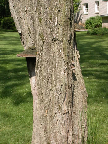
Figure 1. Specimens of Fulvifomes robiniae on black
locust. Photo © Gary Emberger.
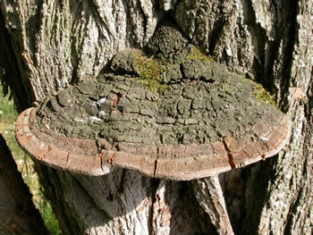
the cracked cap polypore. Photo © Gary Emberger.

Figure 3. The smooth brown pore surface of this
specimen
is 47 cm wide. Photo © Gary Emberger.
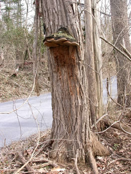
Figure 4. A very large, old specimen.
Photo © Gary Emberger.
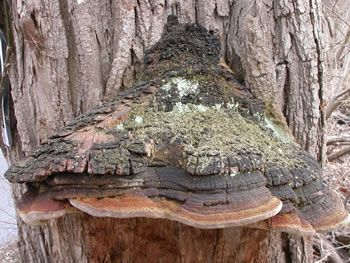
Figure 5. The same specimen pictured in Figure 4 with
moss and lichens growing on
the cap surface.
Photo© Gary Emberger.
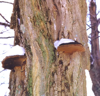
Figure 6. Fulvifomes robiniae is perennial and looks
about the same any time of the year, even with snow on
it. Photo © Larry Grand.
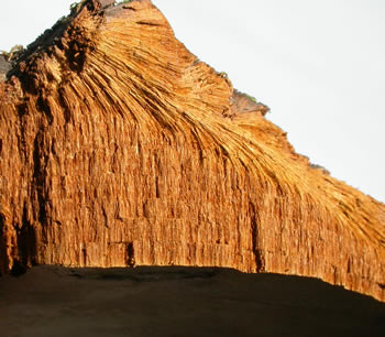
Figure 7. This sectioned specimen shows the layers of
tubes formed by perennial polypores such as Fulvifomes
robiniae. Photo © Gary Emberger.